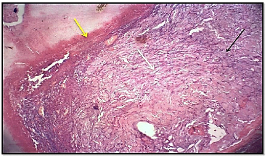
Figure 4
Histological section of wound healing control group at days 7, showed significant highly collagen fibers proliferation ( white arrow). Granulation tissue more cellularity (black arrow) with less presence hemostasis (red blood cells) (yellow arrow). (Hematoxylin and Eosin, 10 X).