
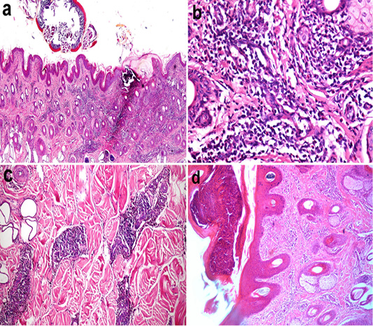
Figure 2
Histopathology of Bovine skin showing (a) tick attached to the surface of the skin with diffuse dermatitis (X40), (b) mononuclear and few eosinophils infiltration in the dermis (X200), (c) panniculitis in the hypodermis, (d) pustule formation in the epidermis (X100). (H and E stain)