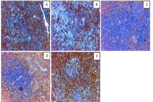
Figure 1
Photomicrograph of mesenteric (paracortex area) lymph node. Positive expression of T-lymphocyte helper receptor (CD4) (arrow) is shown in (A) control group, (B) hypothyroidism group, (C) SC-SeNPs, (D) levothyroxine group, and (E) SC-SeNPs+levothyroxine group. Hematoxylin & DAB. 100x. Note that the lymphoid follicle (arrowhead) in (D and E) did not show CD4 expression.