
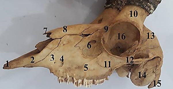
Fig. 6.
Dorso-lateral view of the skull of male chinkara showing incisive bone (1), nasal process of incisive bone (2), maxilla bone (3), infraorbital foramen (4), parietal bone facial tuberosity (5), lacrimal (6), nasal process (7), nasal bone (8), orbital rim (9), cornual process of frontal bone (10), zygomatic bone (11), zygomatic arch (12), temporal fossa (13), tympanic bulle (14), paracondylar process (15) and orbit (16).