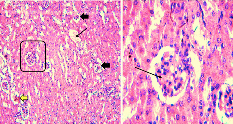
Fig 7
Histological structure of kidney of prednisone treated mouse. Black arrows in (x100) showing affected glomeruli. Not a single glomerulus was normal showing severe lupus nephritis. Thick black arrow in (x400) showing hyaline arteriosclerosis and thin arrow showing glomerular necrosis, brown arrow showing immune deposits of membranous nephropathy, yellow arrow showing tubules with neutrophils.