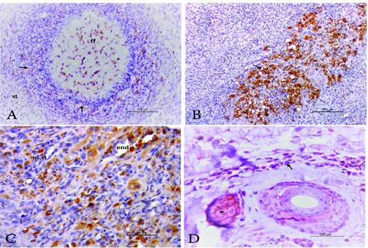
Figure 3
Photomicrograph of ovary and ear notch from PI animal stained by IHC showing, A) growing follicles where the positive cells appear at follicular fluid (ff); granulosa cells (arrow) and at the stromal tissue (st). Stained by IHC. Bar.100µm. B) BVDV antigen positive cells (macrophages) at the atretic follicles (arrow) in the Bar.100µm. C) BVDV antigen positive cells (macrophage) in the ovarian medullar stroma (m.st) as well as endothelium of the blood vessels (end). Bar.50µm. D) ear notch revealed positivity against BVDV antigen in dermal cells (arrow) and sebaceous gland (arrow head); IHC. Bar.100µm.