
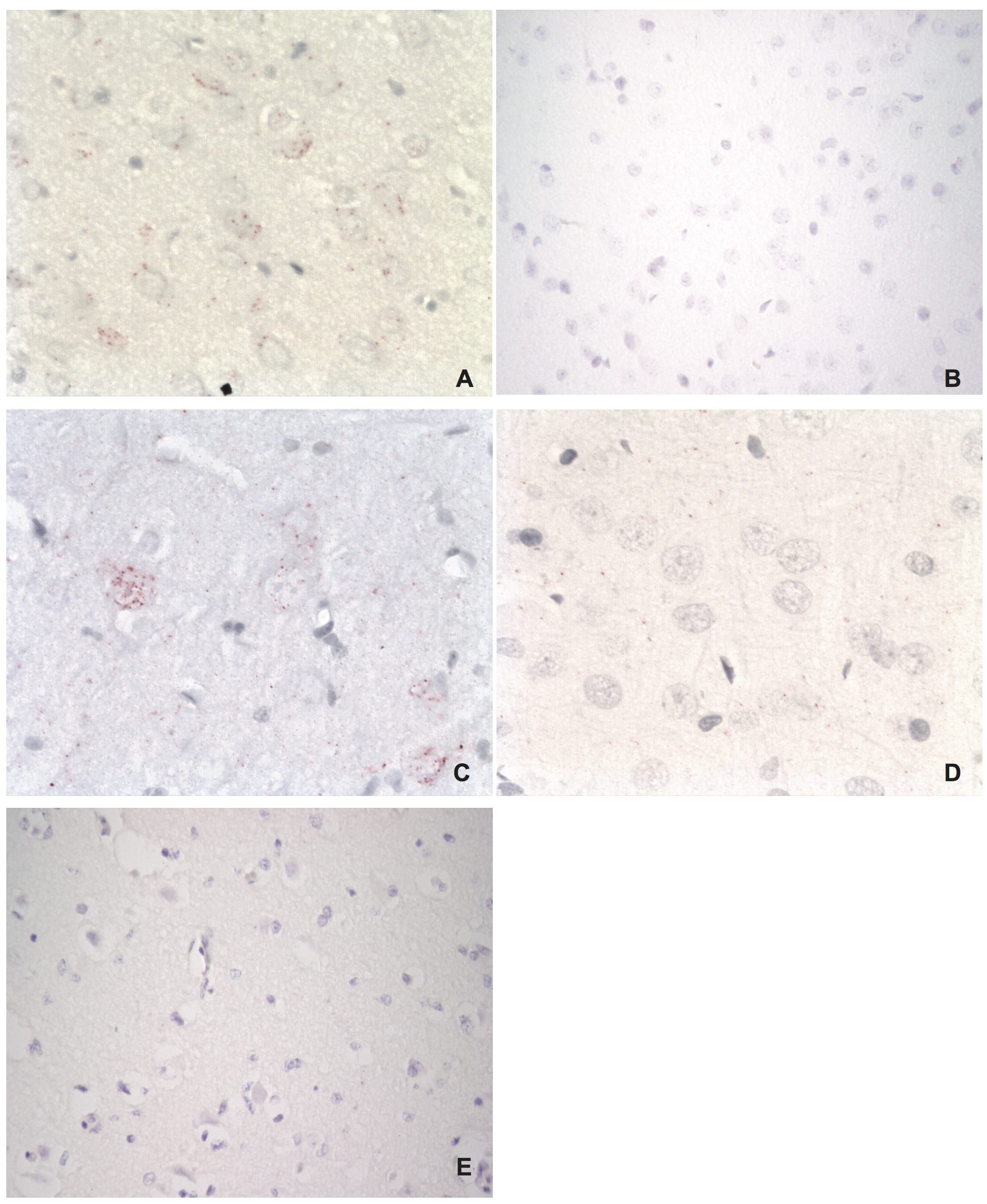
Figure 6
Visualization of host-virus protein-protein interactions in brain tissues of an experimentally infected rat
A) BDV P – HMGB1 interaction (red dots in neuron). B) BDV P – HMGB1 interaction in an uninfected control rat (no signals). C) BDV N – Cdc2 interaction (red dots in neurons). D) BDV N – Cdc2 interaction in an uninfected control rat (no signals). E) Technical control using primary antibody for HMGB1 only in experimentally infected rat. As expected, no signals are seen due to the need for two primary antibodies to obtain a positive in situ PLA reaction. Magnifications: lens x20 (A, C, D), lens x10 (B, E).