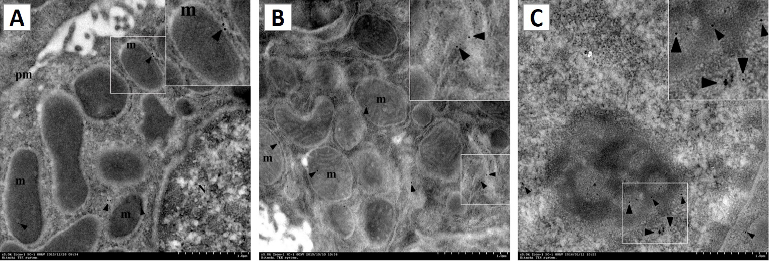
Fig. 4.
Subcellular localization of pGH. Immunoelectron microscopy samples were prepared as described in the “Materials and methods” section. Gold grains were localized to the cell membrane (pm), mitochondria (m) and cell nucleus (N) (A-C). Bar = 1μm. The micrograph represents at least three independent experiments.