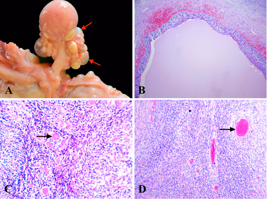
Figure 5
Granulose-theca cell tumor, ovary, camel. (A) Firm grayish-white nodules and numerous yellow cysts (arrow). (B) Macrofollicular pattern with hemorrhage and eosinophilic secretion, HE x 100. (C) Trabecular pattern with Call-Exner bodies (arrow), HE x 400. (D) Diffuse pattern with Call-Exner bodies (arrow), HE x 200.