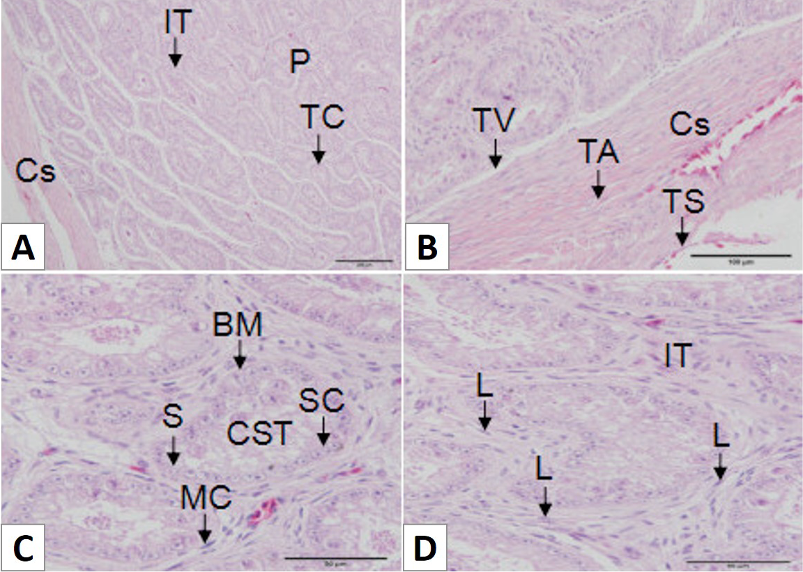
Fig. 1.
Histology of the African Ostrich testes. A, the testis was divided into the capsule (Cs) and parenchyma (P); B, the capsule was divided into three annular layers: tunica serosa (TS), tunica albuginea (TA) and tunica vasculosa (TV); C, the structure of the seminiferous tubules (CST) includes the basement membrane (BM), spermatogonia (S), Sertoli cells (SC) and myoid cells (MC); D, the structure of the interstitial tissue (IT) includes Leydig cells (L), tiny veins and connective tissue. Scale bar: 200 μm (A), scale bar: 100 μm (B) and scale bar: 50 μm (C and D).