
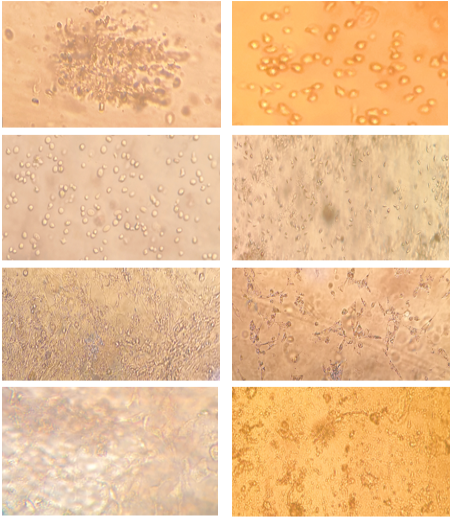
Figure 2
A) First trypsinization, B) second trypsinization, C) third separation shows cell elongation, D) CEFs, E) CEFs maturation, F) CEFs propagation, G) CEFs shows a full cells elongation and forming mono layer, H) CEFs after virus inoculation that from infected CEFs cell, magnification power equal 40x.