Figure 3
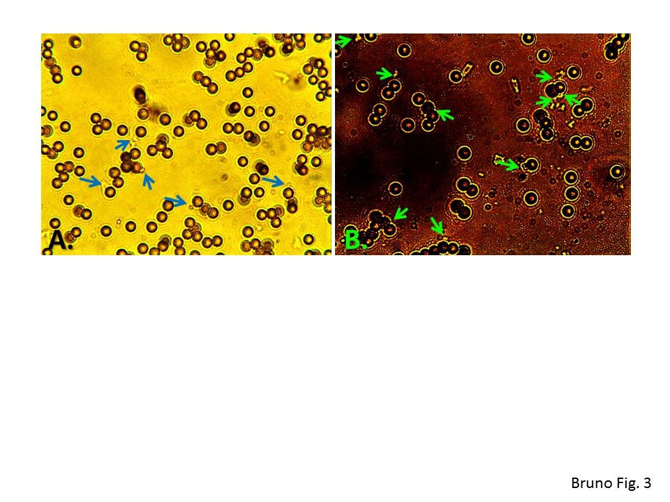
Typical 600X microscopic images of captured acridine orange (AO)-stained Big 7 E. coli bacteria on aptamer-coated magnetic beads (MBs) seen under A) brightfield and B) fluorescence illumination using an FITC (fluorescein) optical cube. Arrows point to captured E. coli bacteria on the MB surfaces
