Fig 1
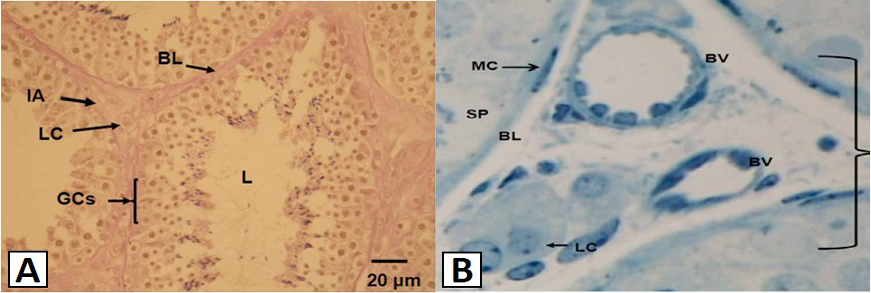
The light micrograph showing the general layout of the interstitial area (IA) and the seminiferous tubules (A), and Leydig cells (LC) and blood vessels (BV) of the testis (B) of the vervet monkey, Chlorocebus aethiops. A, Each tubule is surrounded by a basal lamina (BL). The Leydig cells (LC) are found in the interstitial area (IA). Within the seminiferous tubules are developing germ cells (GCs), which will later become the mature spermatozoa. The lumen (L) is occupied by spermatids of different stages; B, The myoid cells (MC) are found forming the basement membrane or the basal lamina (BL). On the basement membrane lies the spermatogonium (SP).