
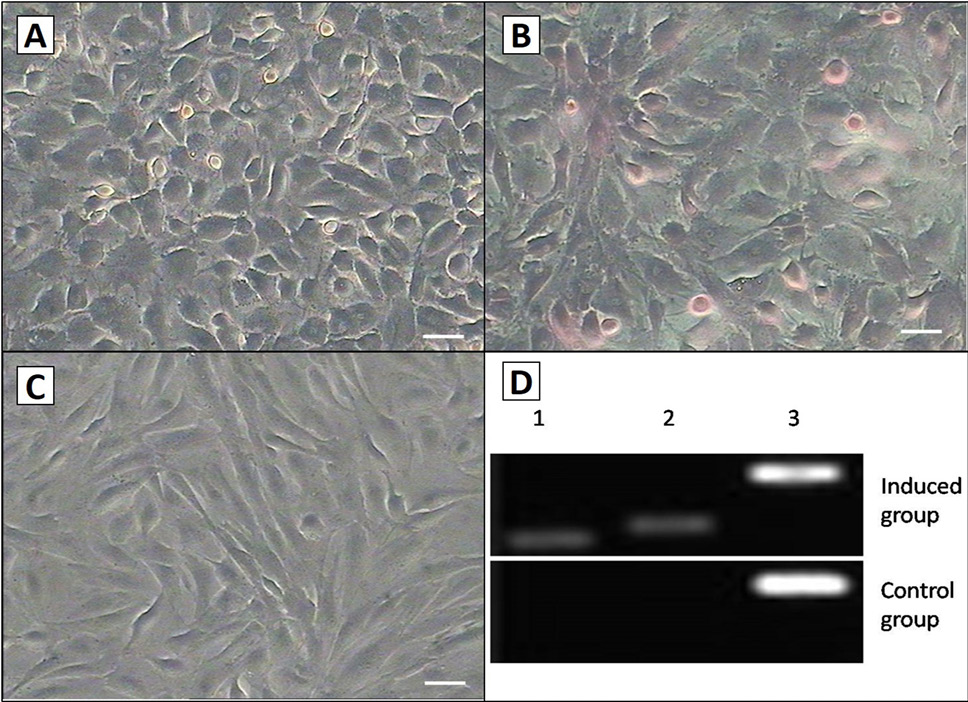
Fig. 8.
Hepatocyte differentiation of the AMSCs. A, after cultured in hepatocyte medium for 14 days; B, the cell shape was changed, and glycogen staining was positive; C, there was no change in morphology or stained by glycogen in control group after 14 d. Scale bar, 50μm; D, RT-PCR analysis of the Hepatocyte differentiation markers ALB and AFP expression in induced group and control group. Induced cells were positive for ALB and AFP, but the control cells were not. Lane 1, ALB; Lane 2, AFP; Lane 3, GAPDH.