
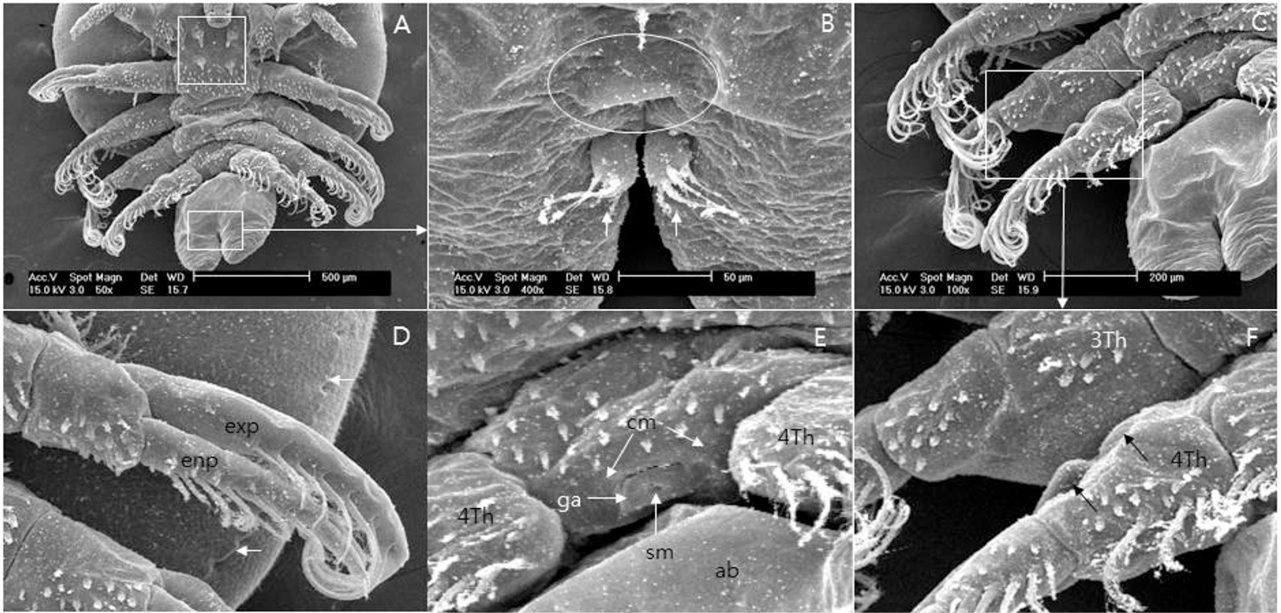
Fig 3
Scanning electron micrographs of postero-ventral view of male A. japonicus. A, Thorax and abdomen. Spines of each post maxillary spine (thoracic spine?) (square); B, Anus (circle). Caudal rami (arrow). Note the four setae; C, Third and fourth leg; D, First swimming leg; Exopodium and endopodium with plumose setae. Posterior region of respiratory area (arrows); E, Male’s genital aperture to show spermatophore secretion; F, Peg (arrow) on the right fourth leg and the socket of right third leg. Note the socket (indentation) on the ventral side of forth leg. ab, abdomen; cm, contracted muscle; exp, exopodium; enp, endopodium; ga, genital aperture; sm, spermatophore; 3Th, third leg; 4Th, fourth leg.