
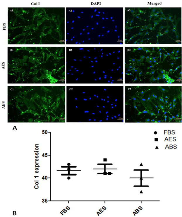
Fig. 2.
A, Expression of Collagen type1 (Col 1) on ATFs after supplementation with 10% FBS (A1-A3), 10%AES (B1-B3) and 10%ABS (C1-C3). Cells were stained with anti-type1 collagen and FITC conjugated secondary antibody whereas DAPI was used to stain the nucleus (X200). B, Expression of Collagen type1 (Col 1) in ATFs cultured with 10% FBS, AES and ABS sera. Scattered dot plot shows that there is same level of Col type1 expression all groups. Data was expressed as Mean ±SEM and level of significance was < 0.05.