
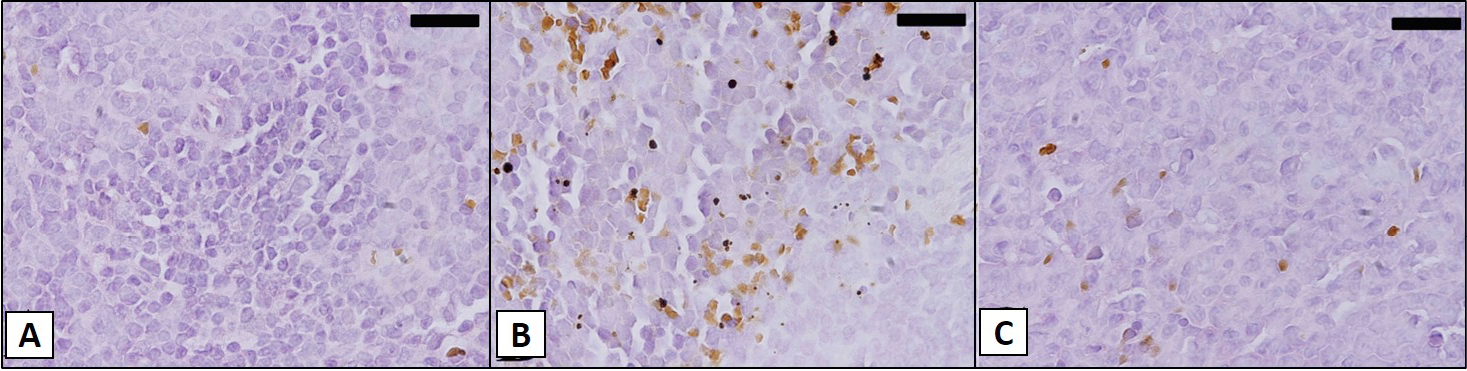
Figure 2
The effect of malarial infection on apoptotic DNA fragmentation in splenic tissue of female BWF1 mice splenic tissues of the lupus (A), live P. chabaudi (B), and gamma-irradiated P. chabaudi (C) infected group of BWF1 mice at week 32. Paraffin embedded tissue sections were prepared and investigated using TUNEL apoptosis detection kits. The TUNEL-positive nuclei are markedly different from those observed in the lupus group. Scale bar = 25µm.