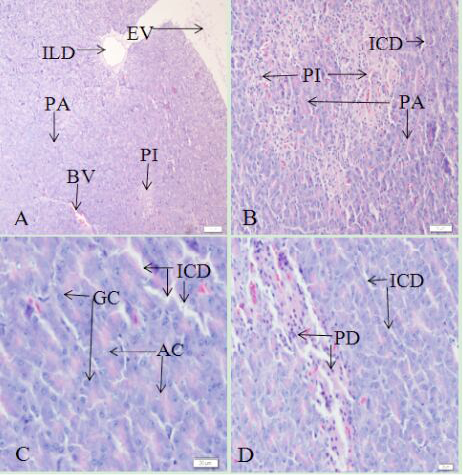
Fig 1
Histological structure of the African Ostrich pancreas (HE staining). A, the pancreas; B, the pancreatic parenchyma; C, the pancreatic exocrine portion; D, the pancreatic parenchyma. EV, envelope; PA, pancreatic acinus; PI, islet; BV, blood vessels; ILD, intralobular duct; ICD, intercalary duct; GC, pancreatic gland cells; AC, centroacinar cell; PD, islet cells. (A) Scale bar: 100 μm; (B) scale bar: 50 μm; and (C and D) scale bar: 20 μm.