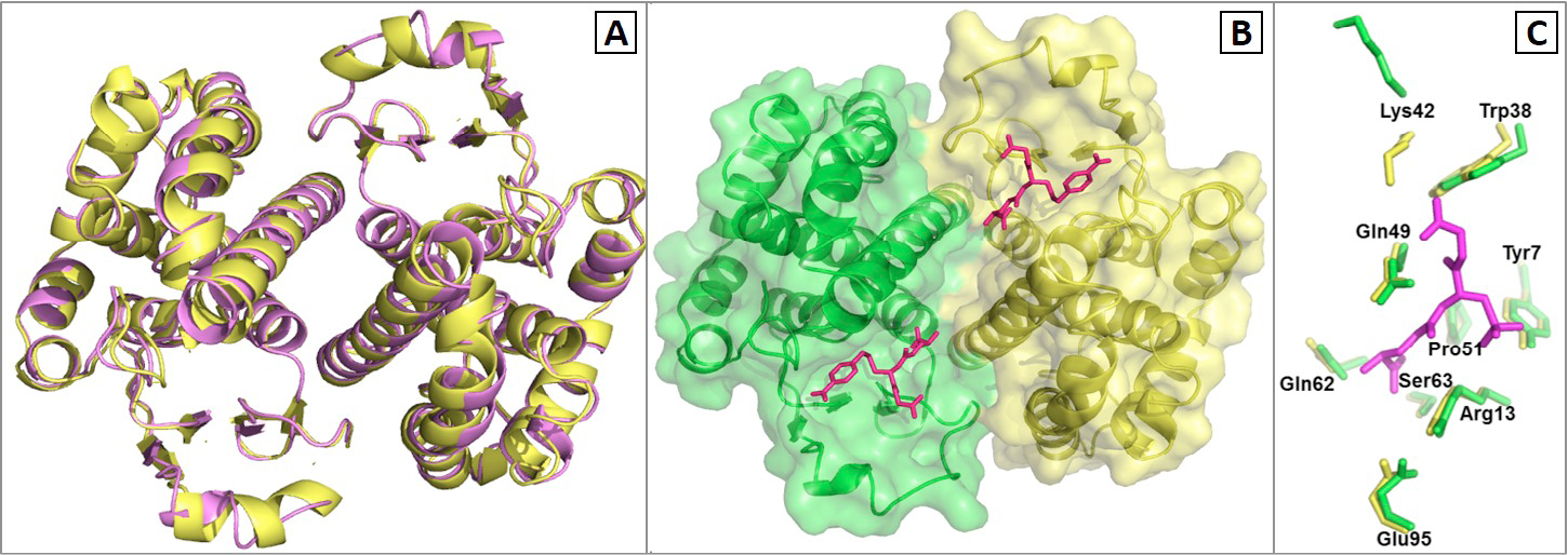
Fig. 7.
A, Superimposed modeled CdGSTP1-1 (violet) on template porcine GSTpi (yellow). The modeled CdGSTP1-1 indicated very high similarity in folding pattern with porcine GSTpi; B, surface view of the G- and H-site of CdGSTP1-1. The two subunits are shown in green and yellow. The bound inhibitor, glutathione sulfonate, is shown with magenta color; C, Comparison of the G-site binding residues and other important residues in camel (green) and porcine GSTpi (yellow).