
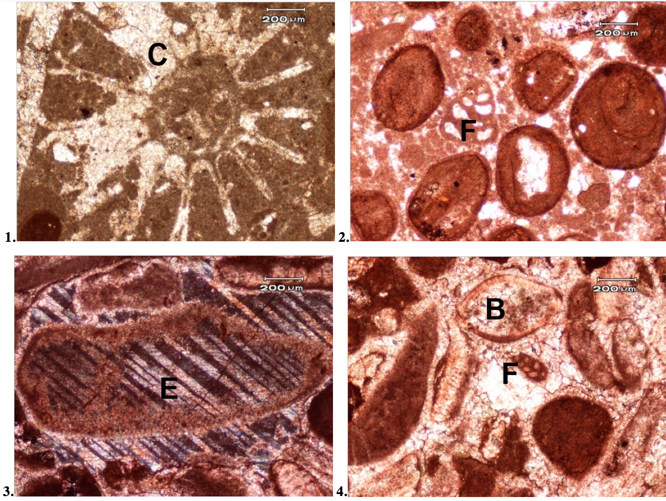
Plate 3:
The photomicrographs showing in bioclastic limestones in Fig. 1: A large coral (C) shell (PPL, unstained), Sample No CHN 61, Fig. 2: A bi-serial (F) foraminifera (PPL, stained), Sample No CHN 61, Fig. 3: An echinoderm (E) grain exhibiting diagnostic single crystal structure extinction (XN, stained), Sample No. CHN-64, and in Fig. 4: Brachiopod (B) and bi-serial foraminifera (F) shells (PPL, stained), Sample No CHN 64