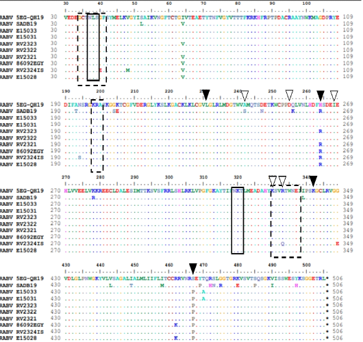
Figure 7
Structural features of G protein. Solid boxes show glycosylation sites. Dashed boxes for antigenic sites II and III. Black arrow heads indicate remaining antigenic sites.White arrowheads indicate pathogenicity related residues.
