
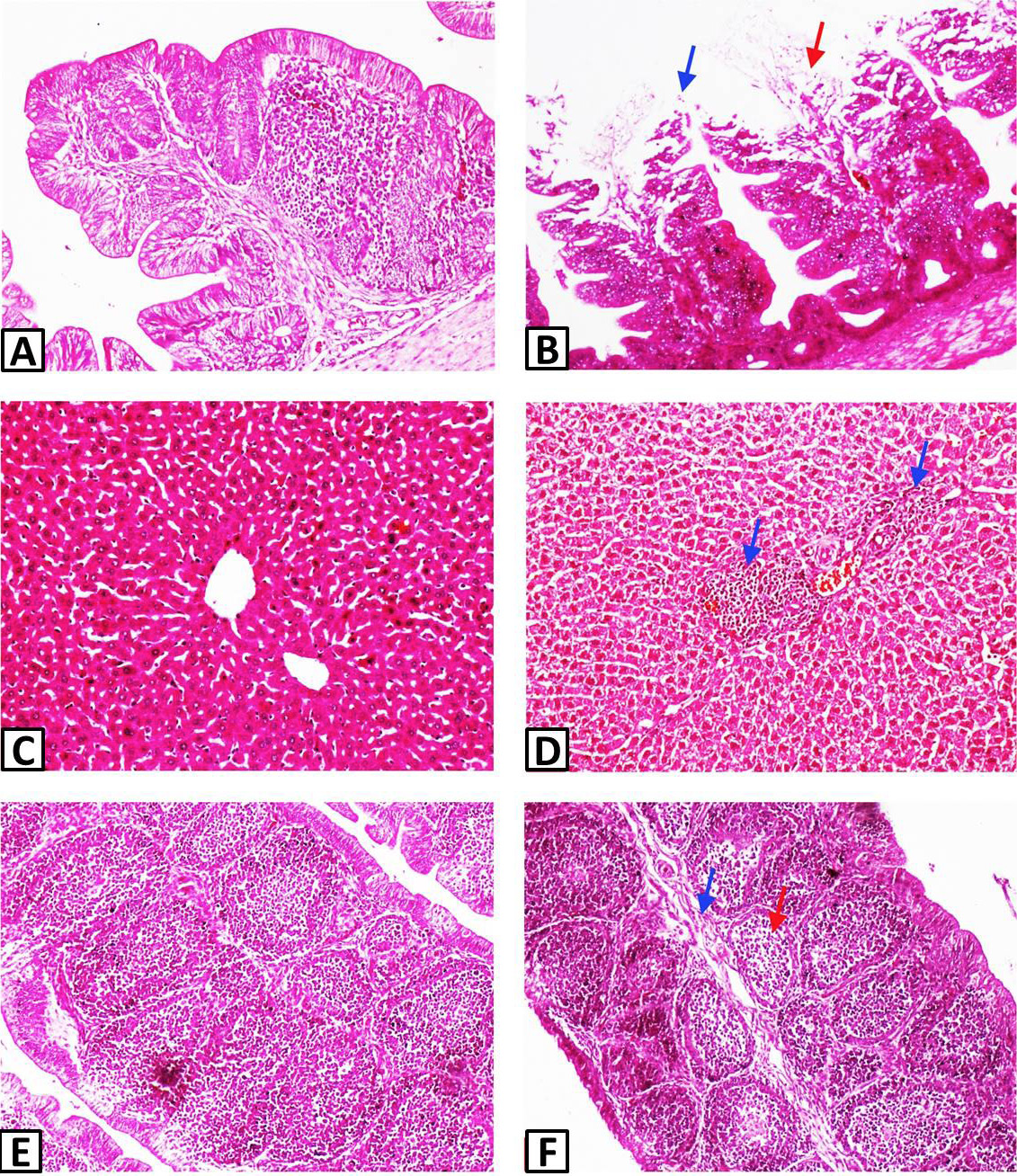
Fig. 1.
Histological structure of parts of different organs of chicken infected with E. tenella. A, normal caeca with intact broad folds. B, necrotic debris in lamina propria (red arrow) and sloughing off epithelial cells of broad folds of caeca of E. tenella infected bird (blue arrow). C, normal liver with intact central vein, hepatocytes and hepatic cords. D, lymphoplasmacytic infiltration in periportal area of liver (blue arrow) of E. tenella infected bird. E, normal BF with intact bursal follicles. F, fibrous connective tissue accumulation (blue arrow) and depletion of lymphocytes in BF (red arrow) of E. tenella infected bird. Stain: H&E; Magnication: 10X.