
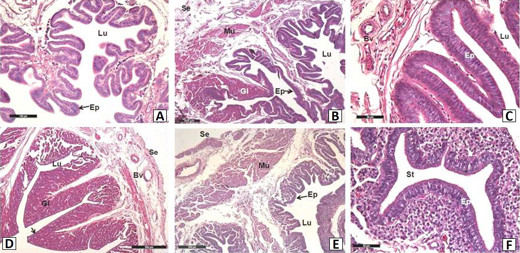
Fig. 1.
Light micrographs in different regions of the hens oviduct. A, infundibulum; B, magnum; C, isthmus; D, uterus; E, vagina; F, the SST in the anterior vagina. Ep, epithelium; Gl, gland; Lu, lumen; St, sperm-storage tubul; Mu, muscle; Bv, blood vessel; Se, serous. Bar=100 μm (A), bar=500 μm (D), bar=200 μm (B, E), bar=50 μm (C, F).