
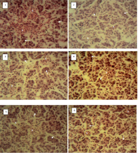
Figure 6
Photomicrograph of liver of C. gariepinus showed normal liver histology with presence of Sinusoid (S); Hepatic cord (arrow) in all the experimental groups before raised in biofloc; Groups 1 (CE), 2 (NE), 3 (CA 10:1), 4 (CA 20:1), 5 (WH 10:1) and 6 (WH 20:1). H&E x400.