
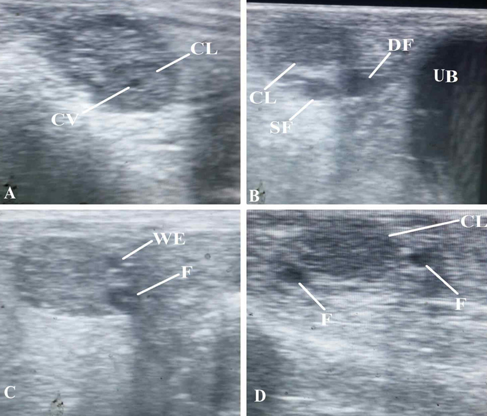
Fig. 4.
Representative ovarian ultrasound images of Lohi sheep. A) Ovary having a corpus luteum (CL) with cavity (CV). B) Ovary detected cranial to the urinary bladder (UB) showing a CL, dominant follicle (DF), and a subordinate follicle (SF). C) Ovary having multiple small follicles (F) at the time of follicular wave emergence (WE). D) Ovary showing mid-luteal phase CL and follicles (F).