
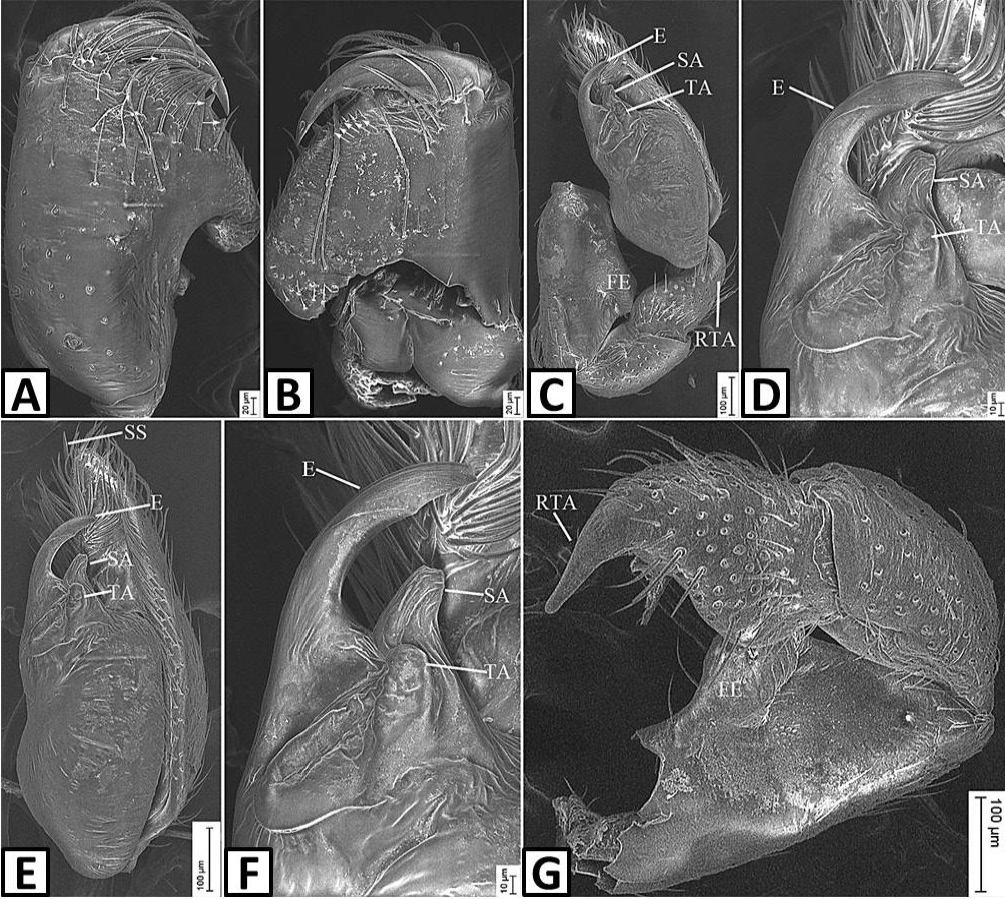
Fig. 3.
SEM micrographs of Otacilia dadongshanica sp. nov., male holotype. A, Chelicera, frontal view, white arrows showing the promarginal teeth; B, Same, posterior view, white arrows showing the retromarginal teeth; C, Palp, retroventral view; D, Same, detail of embolus, subterminal apophysis and terminal apophysis; E, same, retrolateral view; F, Same, detail of embolus, subterminal apophysis and terminal apophysis; G, Same, dorsolateral view, detail of retrolateral tibial apophysis. Abbreviations: E, embolus; FE, femoral extension; RTA, retrolateral tibial apophysis; SA, terminal apophysis; SS, subapical spine; TA, terminal apophysis.