
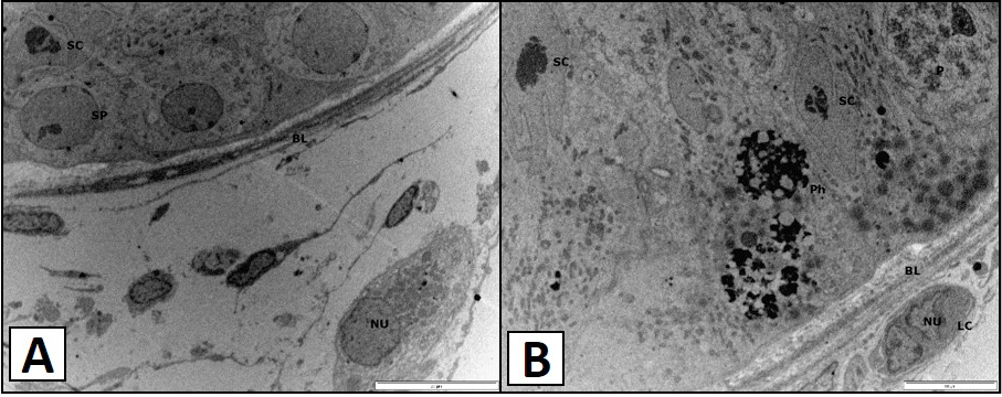
Fig 4
TEM micrograph showing the distinct nucleus (NU) of the Leydig cells of the vervet monkey, Chlorocebus aethiops. A, shows the Sertoli cell (SC) and the spermatogonium type B and found lying on the basal lamina (BL). B, shows the phagocytic vesicles are observed (Ph) and the spermatocyte at the pachytene (P) is observed. The Sertoli cells (SC) are found lying on the basal lamina (BL).