
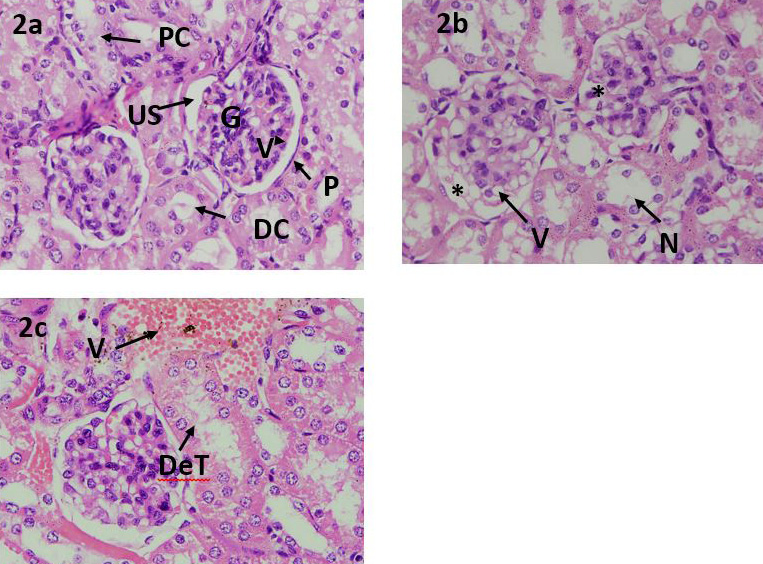
Fig. 2.
(A) Photomicrograph of mice kidney from control group. PCT: proximal convoluted tubules; G: glomerulus; VL: visceral layer; PL: parietal layer; US; urinary space; DCT: distal convoluted tubules, (B & C) Photomicrograph of mice kidney fed with experimental diets. V: vacuolization; N: necrosis; DeT: degenerated tubules; VC: vascular congestion.