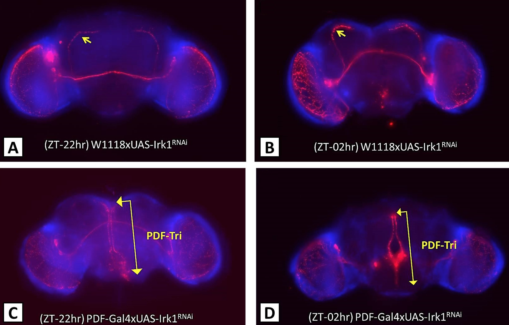
PDF-expressing neurons show a different arborisation pattern in the absence of IRK1 channels. Male flies aged 5-7 days raised on normal food and12:12 LD cycle were beheaded at two different time points. The brains were dissected and stained with anti-PDF antibody (red). A and B, the control flies with sLNvs sending projections towards the dorsal part of the brain (indicated by the yellow arrow) both before and after the lights were turned on. C and D, the IRK1 knock down flies where axonal projections towards the dorsal neurons were absent. The yellow double arrow indicates the presence of PDF-Tri neurons, which are usually degenerated after few days of eclosion. ZT-22 h indicates two hours before the lights were turned on (ZT 0 h = 0800 h), ZT-02 h indicates two hours after the lights were turned on.