
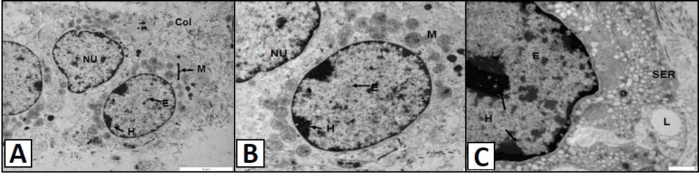
TEM micrograph showing Leydig cells of the vervet monkey, Chlorocebus aethiops. A, shows the polygonal nuclei (NU). The nucleus contains patches of euchromatin (E) and heterochromatin (H), with the latter situated at the periphery of the nucleus, close to the nuclear envelope. The cytoplasm contains numerous mitochondria (M) found closer the nucleus. The collagen fibers (COL) are also observed in the apical region of the cell. B, shows the round nuclei (NU) of the Leydig cells. The euchromatin (E) and heterochromatin (H) are observed in the nucleus. The cytoplasm contains numerous mitochondria (M) found closer the nucleus. C, shows the nucleus and the smooth endoplasmic reticulum (SER) of the Leydig cells. The nucleus contains euchromatin (E) and heterochromatin (H) patches. The lipid droplet (L) is found in association with the SER.