Figure 2:
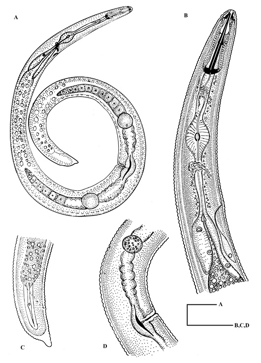
Helicotylenchus fotedariensis sp. nov. A: Entire female; B: Oesophageal region of female; C: Tail region of female; D: Vulval region showing gonad.
Helicotylenchus fotedariensis sp. nov. A: Entire female; B: Oesophageal region of female; C: Tail region of female; D: Vulval region showing gonad.