
Figure 7:
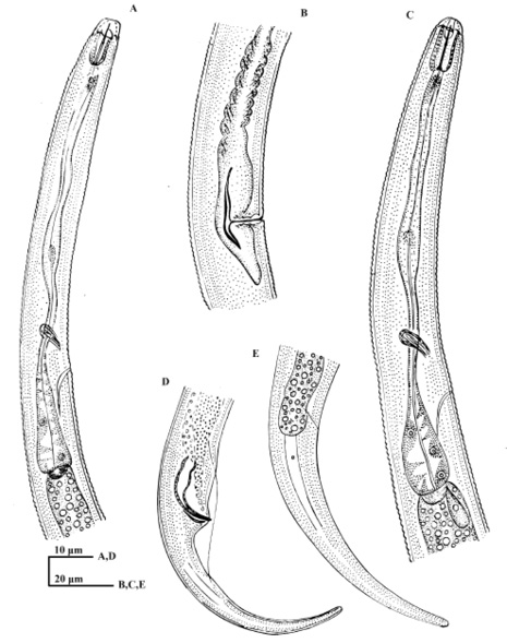
Boleodorus seshadri sp. nov. A: Oesophageal region of male; B: Vulval region showing posterior uterine sac; C: Anterior end of female; D: Tail region of male; E: Tail region of female showing phasmid.
Boleodorus seshadri sp. nov. A: Oesophageal region of male; B: Vulval region showing posterior uterine sac; C: Anterior end of female; D: Tail region of male; E: Tail region of female showing phasmid.