
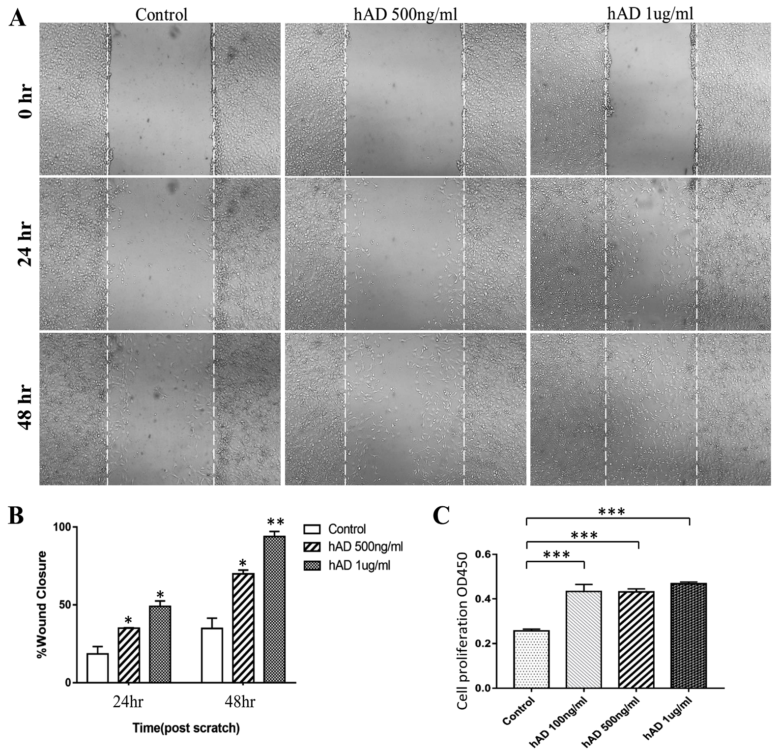
Recombinant hAD promotes NIH3T3 cell proliferation and migration. (A) Representative images from wound healing assays of NIH3T3 cell cultures treated with hAD demonstrating that cell migration into the cell-free region (outlined) is significantly accelerated in the presence of hAD when compared to controls. Images were captured at 24h and 48h after wounding (magnification, ×100). (B) Bar graph illustrating percentage wound closure at indicated time points during the scratch wound assay in the presence of varying concentrations of hAD (*P = 0.040, *P = 0.020, *P = 0.018, **P = 0.008 versus control). (C) NIH3T3 cell proliferation detected using the Cell Counting Kit-8 assay following treatment with hAD (0, 100, 500 and 1000 ng/mL) after 24h (****P<0.0001, versus the control group).