
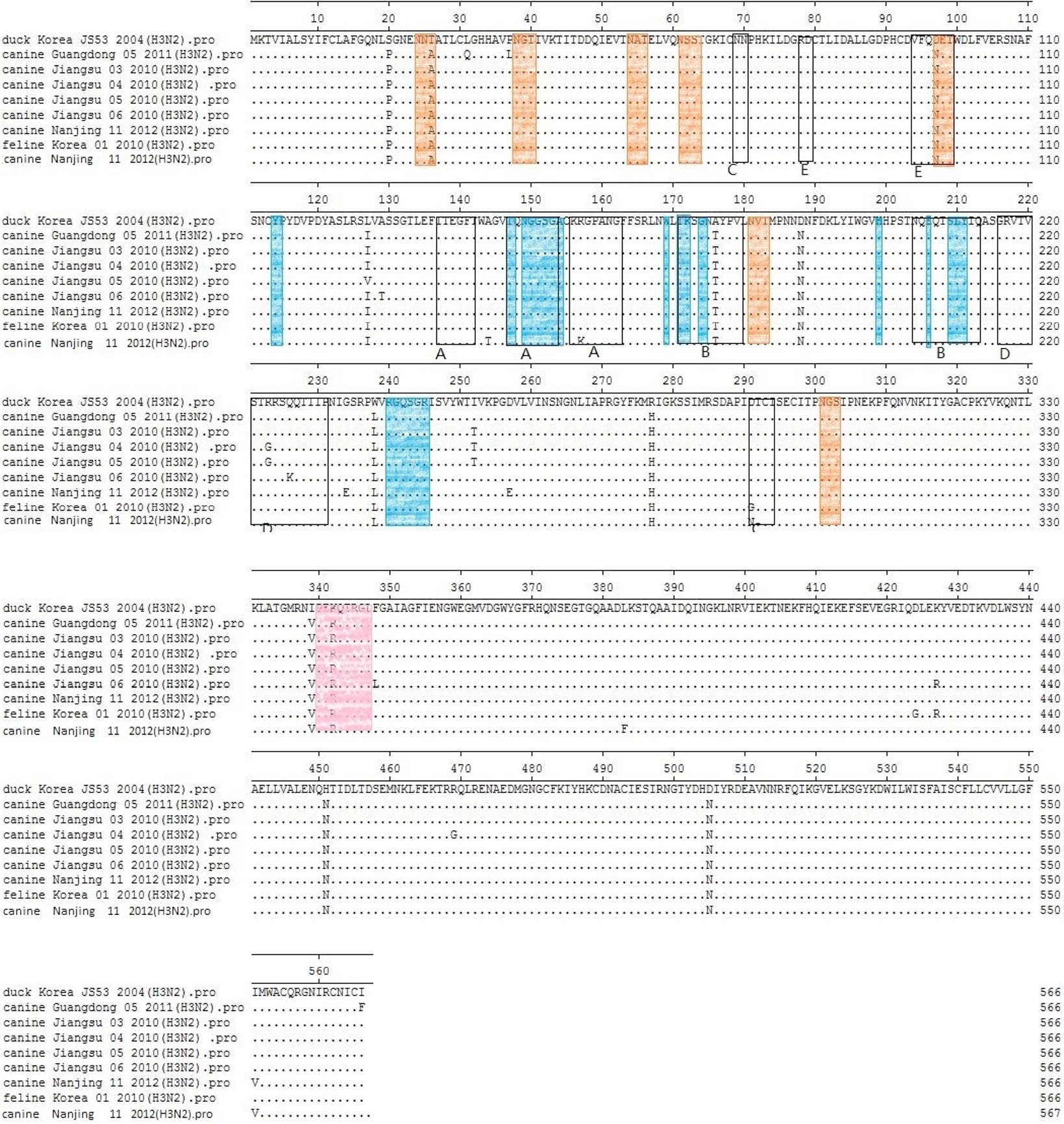
Fig. 5.
Alignment of HA1 amino acid sequences of H3N2 influenza isolates and the most similar avian isolate. Boxed residues represent the antigenic sites A–E, and orange colored residues denote potential glycosylation sites, blue indicates receptor-binding sites and pink represent cleavage site.