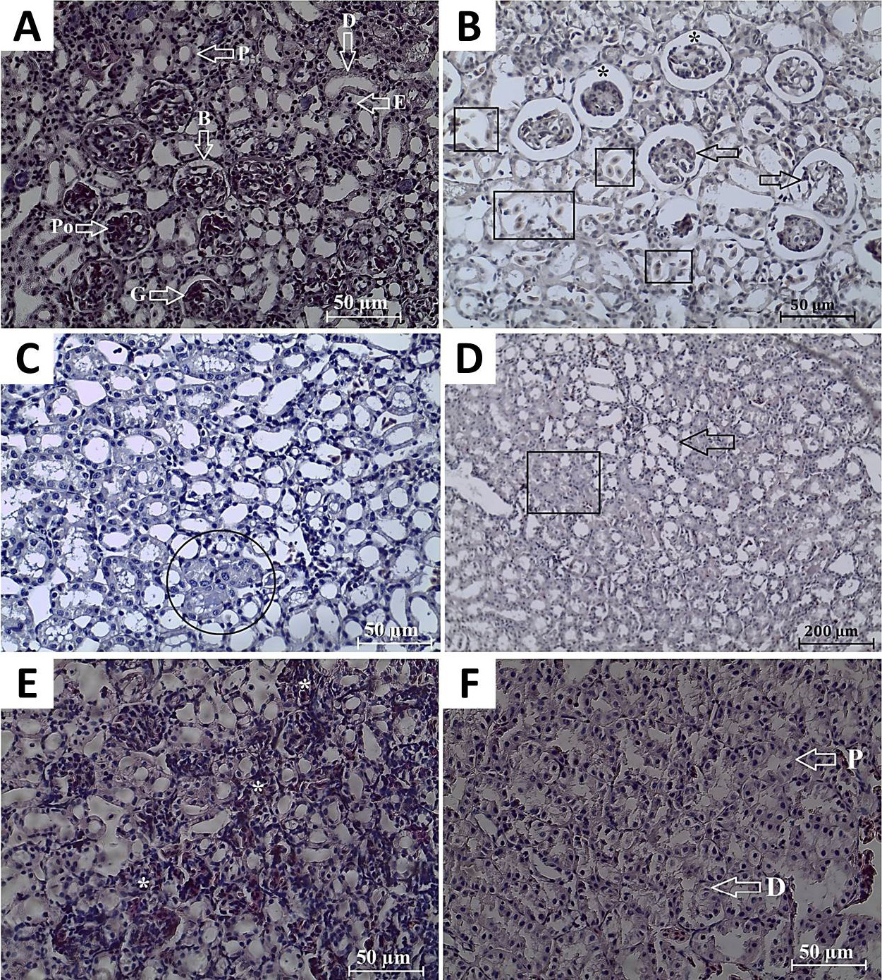
Fig. 4.
A, normal histological properties of the kidneys of P. ridibundus from DB1: renal corpuscles with glomerulus (G) and Bowman’s space (B); podocyte (Po), erythrocytes (E), proximal (P) and distal tubules (D). B, C and D; histological alterations of the specimens of DB2. B, glomerulonephritis (black arrows), congestion in renal parenchyma (squared), and expansion of Bowman’s space (asterisks). C, tubular necrosis (encircled). D, tubular dilatation (arrow), tubule lumens as eosinophilic in appereance (squared). E, F; Histological changes in the kidney of SV specimens. E, lymphocytes infiltration (asterisks). F, degeneration of proximal (P) and distal (D) tubules, (H&E).