
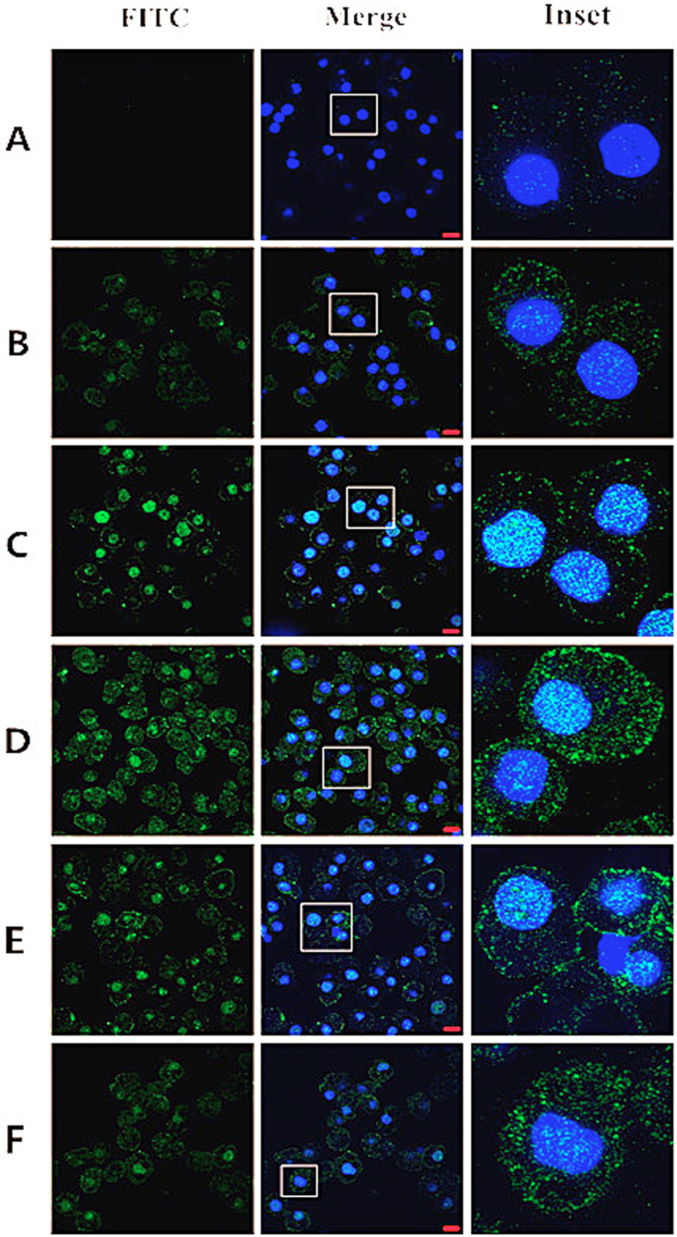
Fig. 2.
CLSM analysis to observe the internalization and nuclear localization of pGH in porcine hepatocytes. Porcine hepatocytes were serum starved, washed and incubated with FITC-pGH (100 ng/mL) for 0, 15, 30, 60, 75 and 90 min and subsequently fixed with 4% paraformaldehyde and stained with Hoechst 33258 (nucleus) (A-F). The cell samples were visualized by confocal laser scanning microscopy. Bar = 10 μm. The images represent at least three independent experiments.