
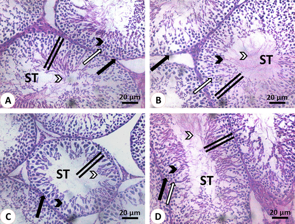
Histological structure of the testis stained with hematoxylin and eosin. (A, B): 3 months old rats before and after quercetin administration, respectively. The seminiferous tubules (ST) showed normal seminiferous epithelium (double line) that contains spermatogonia (black arrow), spermatocytes (white arrow), spermatids (black arrowhead) and mature sperm (white arrowhead). (C, D): The seminiferous tubules (ST) of 30 months old rats before quercetin administration in C showed the depletion of spermatogenic cells (black arrow), round spermatids (black arrowhead) and sperms (white arrowhead). Note the reduction in the seminiferous tubules diameter and its epithelium height (double line). The seminiferous tubules (ST) of 30 months old rats after quercetin administration in D restore the normal structure. Note the height of its epithelium (line), spermatogonia (black arrow), spermatocytes (white arrow), spermatids (black arrowhead) and mature sperm (white arrowhead).