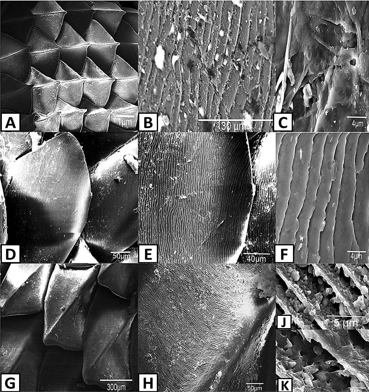
Scanning electron photomicrographs of scale ornamentation in lizards. A, shows the keeled scales of mid-dorsal trunk skin of Acanthodactylus opheodurus (15x); B, shows the pits on the outer surface of mid-dorsal trunk skin scales of A. opheodurus (3000x); C, shows indefinite structures, hair-like and papilla on the outer surface of mid-dorsal trunk skin scales of A. opheodurus (10000x); D, shows the outer surface of mid-dorsal trunk skin scales of Mesalina guttulata guttulata (500x); E, shows the outer surface of mid-dorsal trunk skin scales of M. guttulata guttulata (1000x); F, shows the minute pits on the outer surface of mid-dorsal trunk skin scales of M. guttulata guttulata (10000x); G, shows the keeled scale of mid-dorsal trunk skin of Trapelus ruderatus blanfordi (150x); H, shows the outer surface of mid-dorsal trunk skin scales of T. ruderatus blanfordi (500x); J, shows the microstructure of mid-dorsal trunk skin scales of T. ruderatus blanfordi (5000x); K, shows the microstructure of mid-dorsal trunk skin scales of T. ruderatus blanfordi (30000x).