
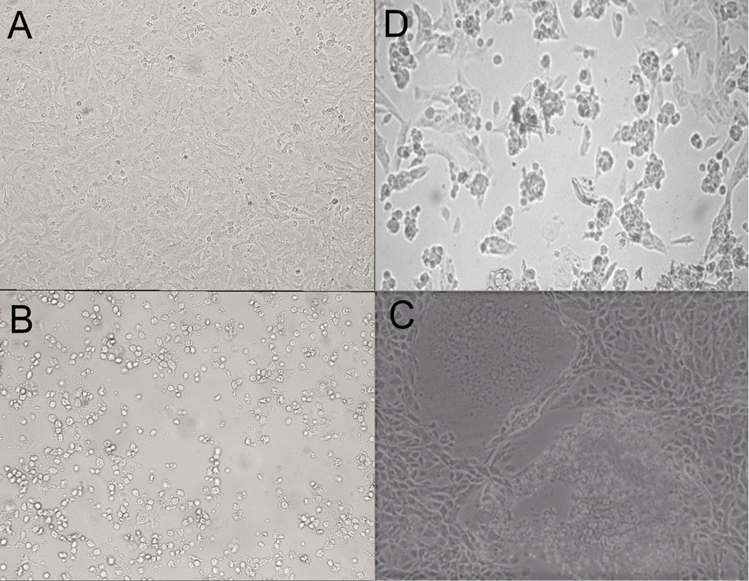
Figure 3:
Microscopic pictures for BCoV CPE in Vero cells (3rd passage) at 100X magnification power; image A: control uninfected Vero cells, image B: rounding of cells 24 H.P.I, granulation, image C: swelling and syncytium formation 96 H.P.I., image D: cell lysis and detachment 5 days post-inoculation.