
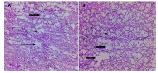
Figure 8
Micrograph of the longitudinal section of control group at the site of the spinal cord injuries A. 8 weeks PO shows multiple cystic cavity, containing granular cellular debris (thin arrows) surrounded by reactive gliosis and presented debris of necrotic with prominent vacuolization in white matter (thick arrow). B. 16 wks shows large cystic cavity (thick arrows) surrounded by glial scar tissue and vacuolated nerve fibers (thin arrow) H&EX10.