
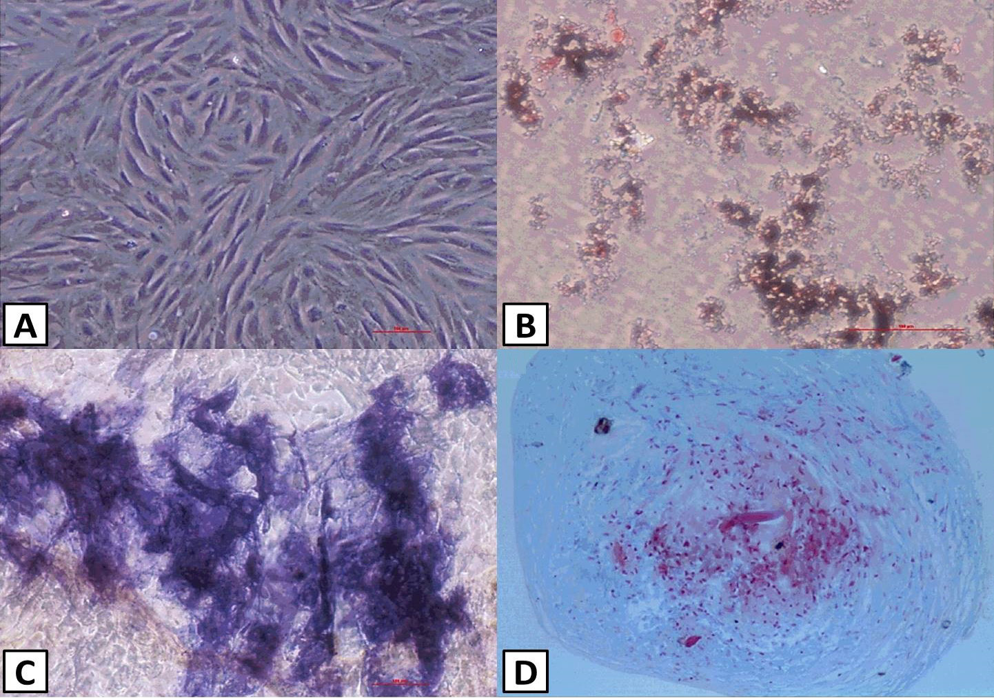
Fig. 1.
Identification of uMSCs. uMSCs exhibited a spindle- and fibroblast-like shape (A). B-D shows multipotential differentiation of uMSCs. uMSC differentiation into adipocytes, osteoblasts and chondroblasts, as shown by Oil Red O (B), alkaline phosphatase (C), and alcian blue (D) staining of in vitro differentiation cultures, respectively.