
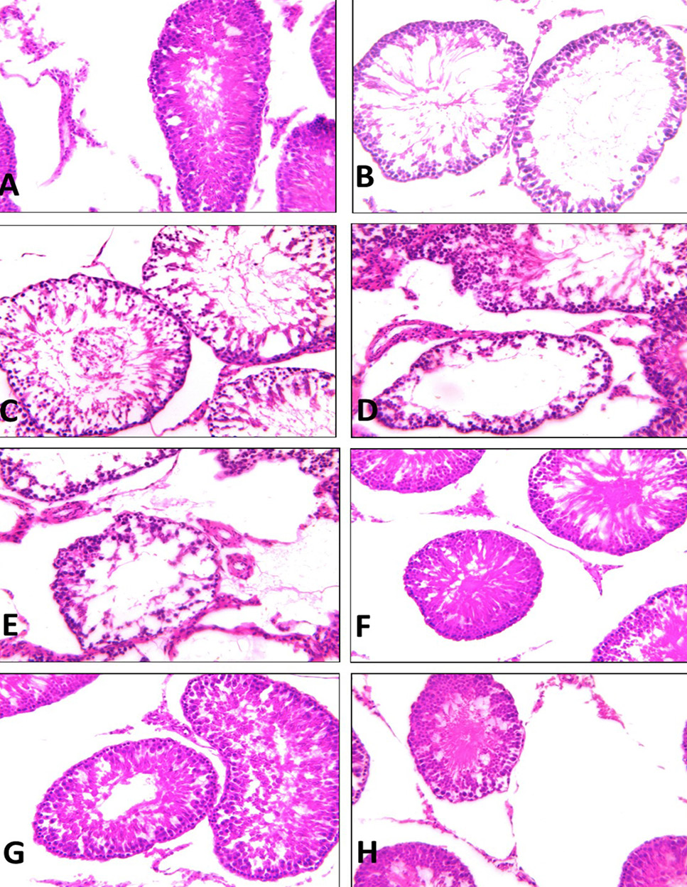
(A) Testis of control rats showing the normal histological structure of seminiferous tubules with different series of active spermatogenic layers, spermatozoa and the interstitial tissues. (B) Testis of mint treated group showing alternations of some tubules in the form of reduced the number of layers of the germinal epithelium, spermatozoa with vacuolated spermatogenic cells. (C, D and E) Testis of AL, AM and AH group showing tubular shrinkage with extensive degeneration of the germinal epithelium. The shrinkage tubules contained degenerated Sertoli cells with few germ cells. (F, G and H) Testis of ALM, AMM and AHM group showing no prominent histological changes. Most of the seminiferous tubules appeared to increase of spermatogenic cells and an increase in the number of sperm bundles was seen. (Stain HX and magnification x200).