
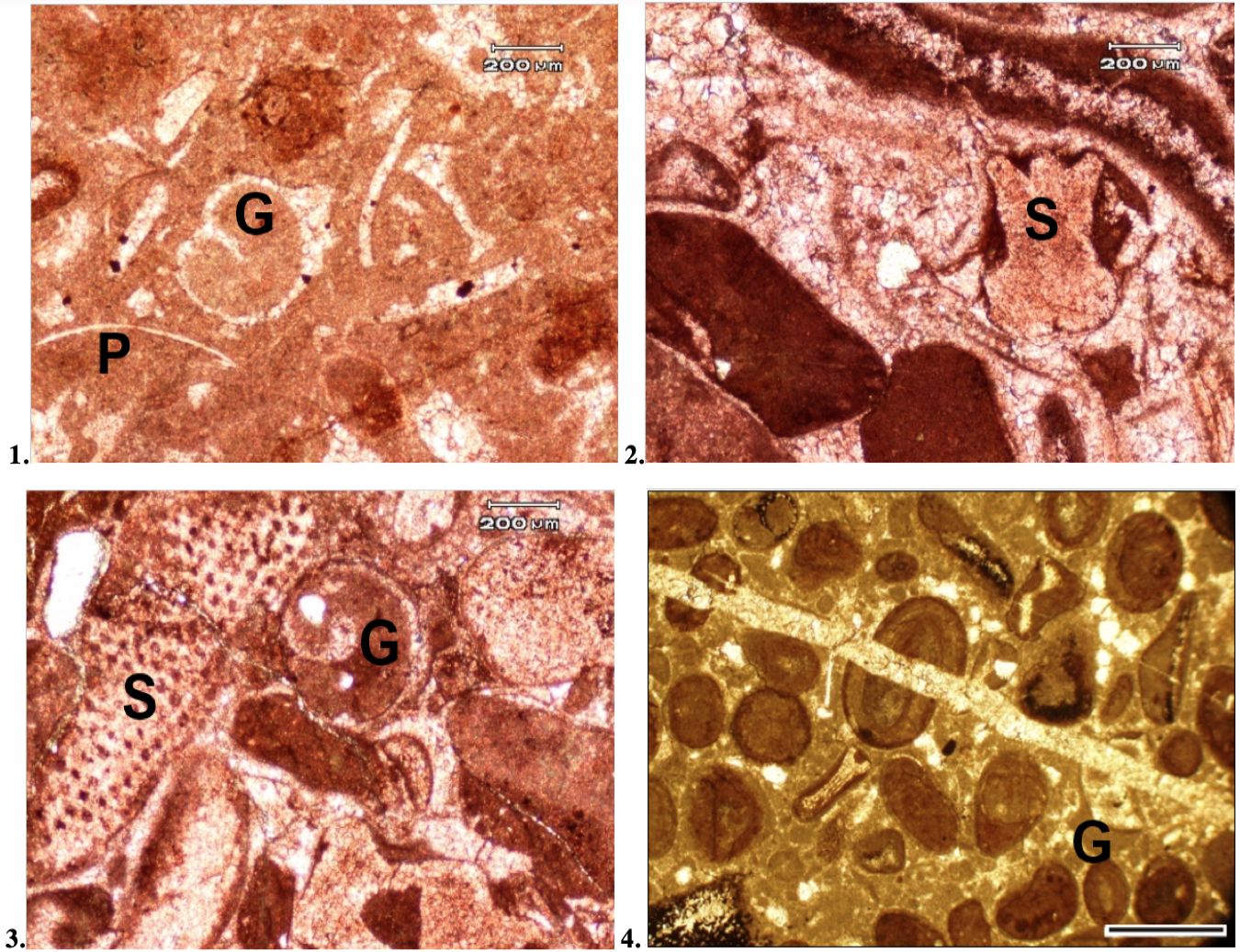
Plate 2:
The photomicrographs showing in bioclastic limestones in Fig. 1: Gastropod (G) shell and pelecypod (P) fragments (PPL, stained), Sample No. CHN 38, Fig. 2: Sponge (S) shells (PPL, stained), Sample No. CHN 39, Fig. 3: Sponge (S) and gastropod (G) shells (PPL, stained), Sample No. CHN 60, and in Fig. 4: A gastropod (G) shell in an ooidal limestone (PPL, stained, Scale bar=250 µm), Sample No. CHN-61