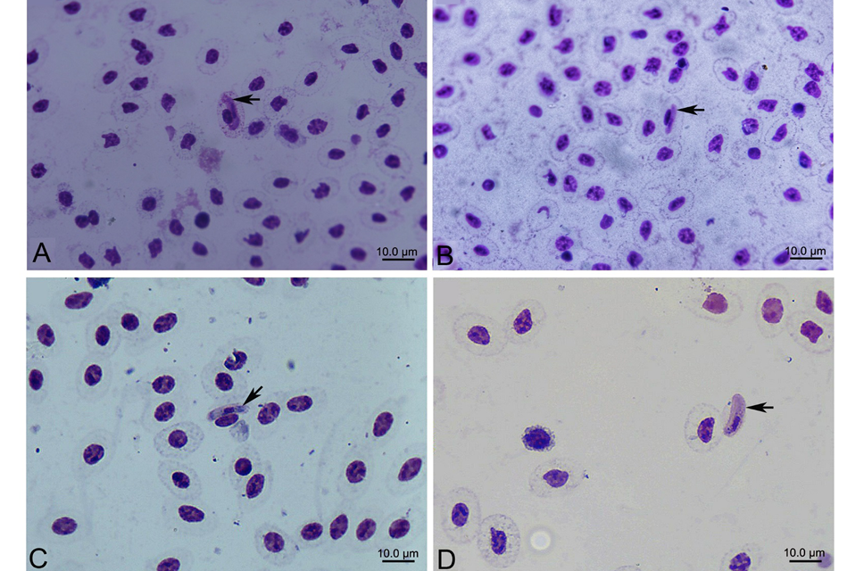
Hepatozoon sp. gamonts from Elaphe carinata and Naja atra. Blood smears stained by Giemsa (×1000). (A) the gamont folded back hook-wise at one end, nucleus of the gamont located in the central, infected erythrocytes became slightly hypertrophic; (B) nucleus of the gamont extended into the quarter, nucleus of infected erythrocytes was flatter than uninfected erythrocytes and infected erythrocytes became slightly hypertrophic; (C) the gamont’s both ends were obtuse and bend slightly to one side in the shape of a kidney, nucleus of the gamont located in the center, nucleus of infected erythrocytes was forced to one side of the host cell and flatter than in the uninfected erythrocytes; (D) gamont was outside of the erythrocyte and nucleus of the gamont extended into the quarter. Arrow indicates gamont of Hepatozoon sp.