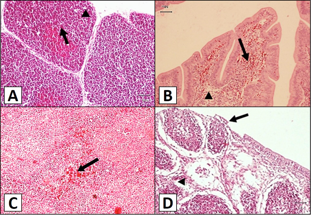
Fig 1
Histological structure of chicken thymus, bursa and spleen. A, thymus of group A at 3rd day post-infection (PI) showing hemorrhages (arrow) and mild lymphocytic depletion (arrowhead) in the follicles; B, Bursa of group A at 5th day PI showing severe hemorrhages (arrow) and lymphocytic depletion (arrowhead) in bursal follicles. C, spleen of group B at 5th day of PI showing severe congestion (arrow) and leukocytic infiltration (arrowhead); D, bursa from group A at 9th day of PI showing severe lymphoid follicle necrosis (arrowhead) with degraded epithelium (arrow).