
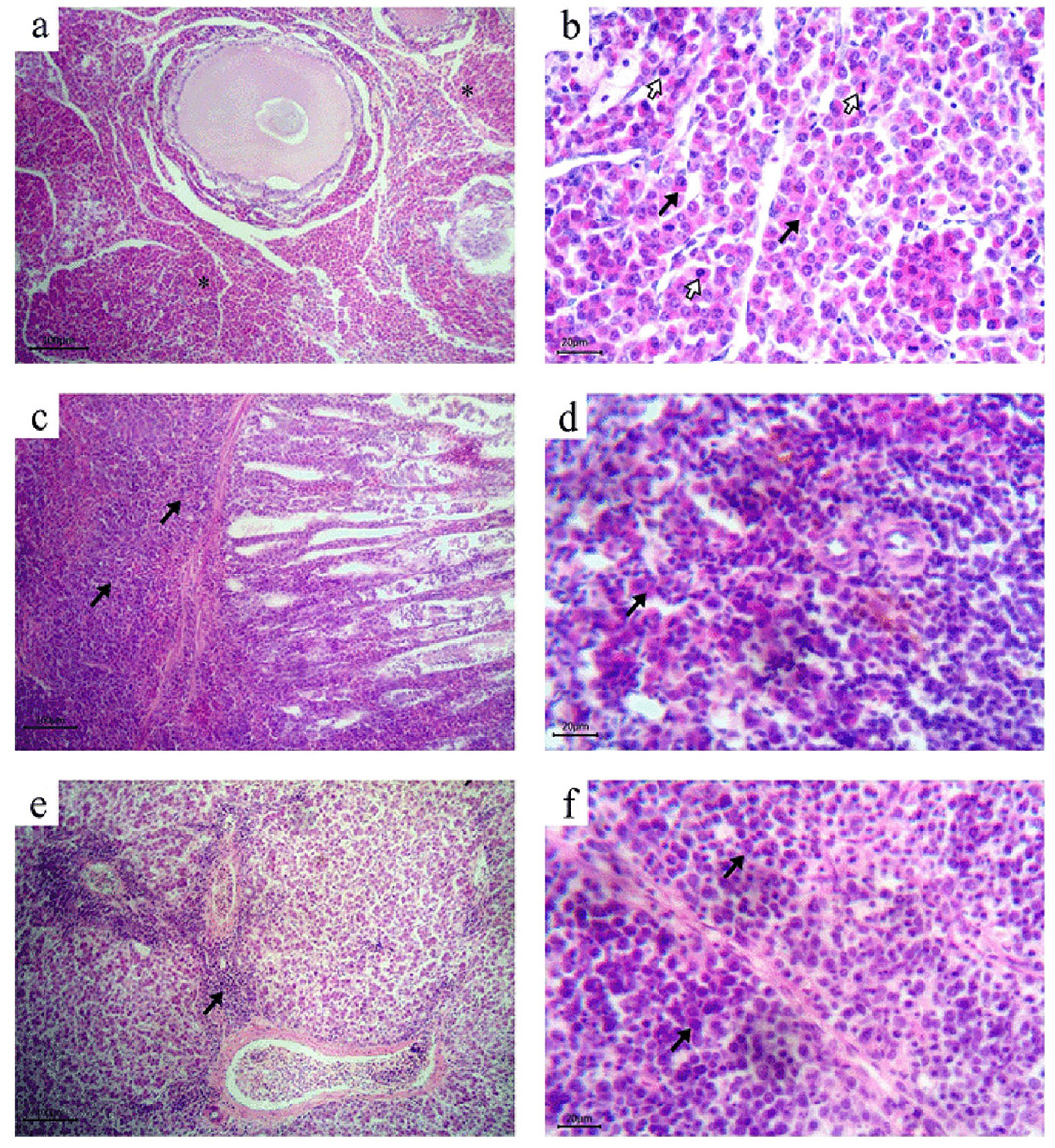
Figure 1
Histopathology of MDV naturally infected birds (HandE stain). a): Ovary. Massive diffuse infiltration with myelocytes (asterisks) with its characteristic eosinophilic cytoplasm; b): Ovary. Myeloid cells proliferation (black arrows) with numerous mitotic figures (white arrows); c): Proventriculus. Diffuse infiltration with neoplastic lymphoid cells both in the lamina propria (arrows) and the proventricular glands; d): Spleen. Mixed population of lymphoid tumour cells and myeloid cells (arrow) adjacent to the central arterioles; e): Liver. Periportal infiltrations with neoplastic lymphoid cells (arrow); f): Bursa of Fabricius. Diffuse infiltrations with pleomorphic neoplastic lymphoid cells (arrows).