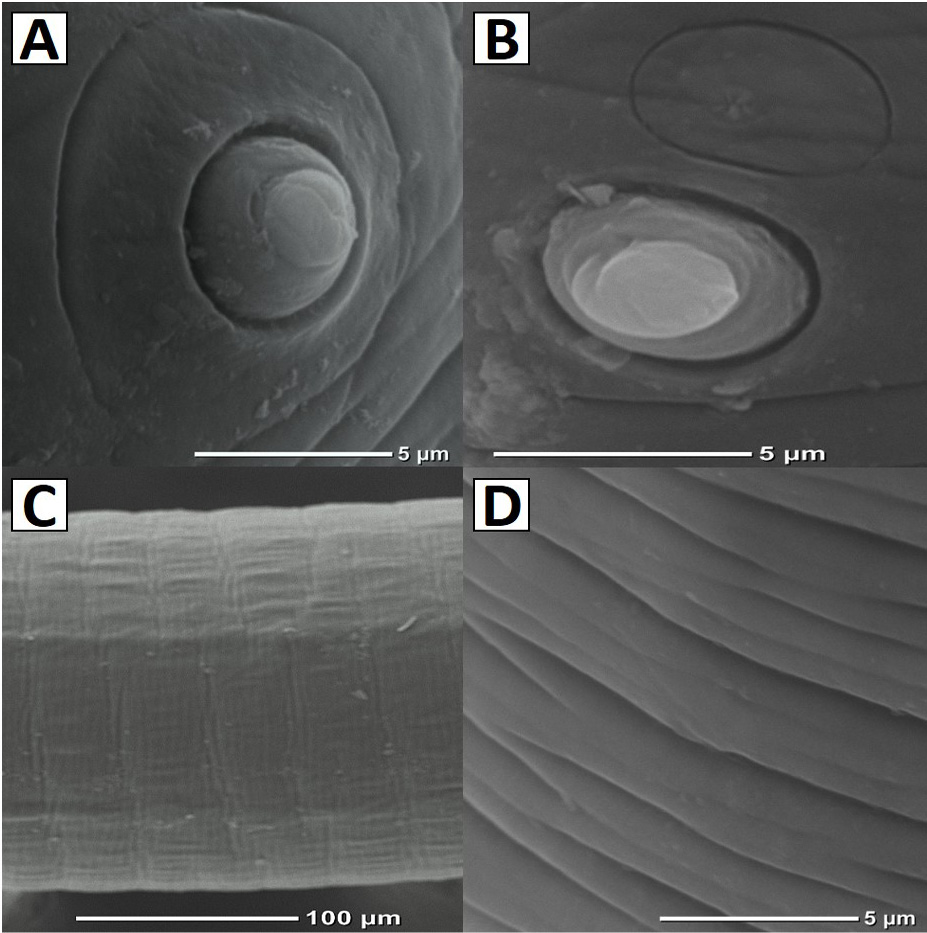
Fig. 5.
Scanning electron micrographs of L4 Eustrongylides tubifex: A, surface view of outer papilla encircled in a ring; B, sensillae situated above the outer papilla; C, body showing striations; D, striations under higher magnification (note the grooves and incomplete margins).