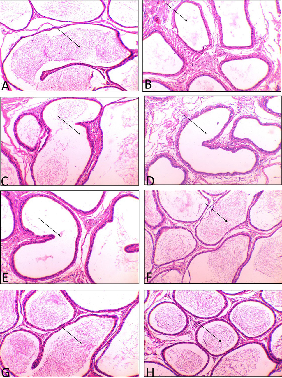
Fig. 4.
(A) Tubuli recti of the control group showing the normal histological structure lining with cuboidal epithelium and present of sperm in the lumen (←). (B) Tubuli recti of mint treated group showing alternations of some tubules in the form of reduced sperm in the lumen (←). (C, D and E) Tubuli recti of AL, AM and AH group showing vacuolation of the cuboidal epithelium with extensive absent of the sperm in the lumen (←). (F, G and H) Tubuli recti of ALM, AMM and AHM group showing no prominent histological changes with increase of sperm in the lumen (←). (H and E, x200).