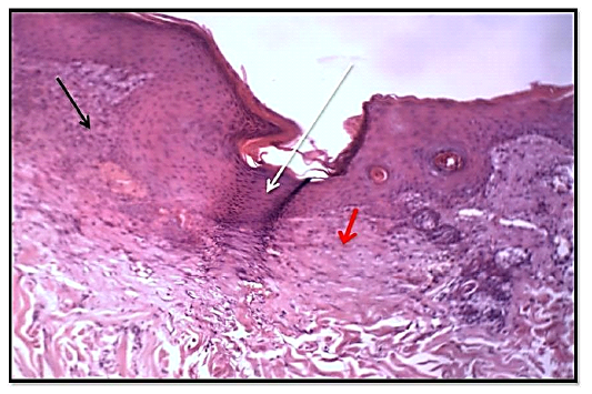
Figure 7
Histological section of 14thday wound healing control group showed the fully epithelized surfaces (white arrow), less collagen fibers proliferation (red arrow). Granulation tissue less cellularity (black arrow) and tissue structure resemble normal. (Hematoxylin and Eosin, 10 X).