
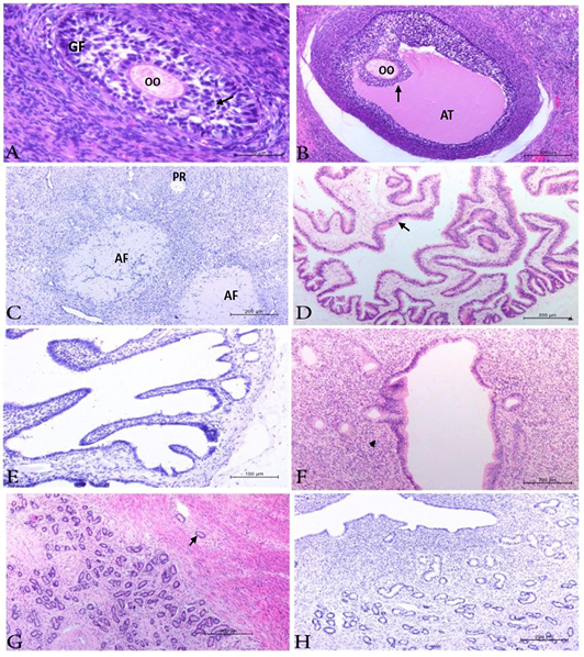
Photomicrograph from ovary, uterine tube and uterus of negative cows showing, A) ovary with growing follicles (GF) had intact oocyte(OO) and corona radiata (arrow). H&E. Bar.50µm. B) ovary with mature follicles (MF) had intact oocyte (OO) and corona radiata (arrow) as well as antrum (AT). H&E. Bar.200µm. C) Ovary with primordial (PR) and collapsed atretic follicles (AF). No positive reaction could be detected. IHC. Bar.200µm. D) uterine tubes showing normal mucosa and sub mucosa. H&E. Bar.200µm. E) uterine tubes appear free from BVDV antigen. labelled by IHC Bar.100 µm. F) Uterus showing mild chronic endometritis (arrow head). H&E. Bar .200 µm. G) Uterus with endometrial gland hyperplasia associated with endometriosis (arrow). H&E. Bar.200µm. H) Uterus showing negative BVDV; antigen. IHC. Bar.200 µm.